| Core Laboratories |
|
Cardiovascular Core Laboratories are academic cardiovascular imaging Core Labs which provide internationally recognized expertise in efficient and environment. Core Labs provide the result of an unbiased interpretation about pharmaceutical or mechanical intervention in coronary artery disease and cardiac transplant studies. The independent processes reduce inter-observer or intra-observer variability and increase the accuracy and precision of results. Also Core Labs ensure that the investigators are adequately trained to optimize the yield of the imaging data and to figure out that all regulatory requirements are in place. It also assists with data analysis and interpretation, presentations to the medical community and education of the regulatory agencies about the study results.
|
| The tasks of the Core Laboratories |
- GCP guideline compliant
- Protocol design of phase III and IV studies
- Case report form design
- Data collection and Interpretation
- Imaging analysis of angiography and intravascular ultrasound
- Data report
- Data analysis collaborated with data management center
- Education for physicians, technicians and nurses
- Collaborations with important core laboratories and data centers
|
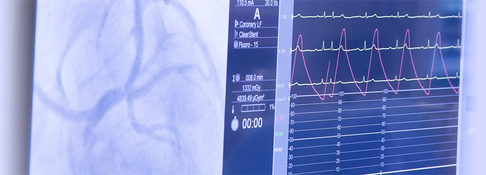
| Quantitative Coronary Angiographic (QCA) Core Laboratory |
|
QCA Core Lab exploits and adjusts catheterization and other procedures for imaging protocols for each clinical trial and project. It achieves perfect and elaborate analysis of data for patients and clinical trial research. These accomplishments are based on ceaseless attempts, aiming for perfection. QCA Core Lab has experience in providing analysis for clinical studies of percutaneous coronary or peripheral interventions with stent, angioplasty, new coronary devices, and interventions with concomitant drug therapy. Importantly, QCA Core Lab is dedicated to the collection of data on TIMI grading and frame counting, morphology assessments for AHA/ACC lesion classification, QCA, including minimum lesion diameter, maximum percent diameter and area stenosis, normal (reference) vessel diameters and lesion length, left-ventricular ejection fraction and regional wall motion results, and ventricular volume indices. Together with interdisciplinary collaborations and consultations, QCA Core Lab provides unique and timely opportunities in interpreting the results of clinical trials and managing clinical researches.
|
| Facilities |
- Research laboratory room
- Plain QCA software: Pie Medical Corp, CASS-5
- Dedicated bifurcation angiographic analysis software
- Angiographers
|
| Intravascular Ultrasound (IVUS) Core Laboratory |
|
IVUS Core Lab offers a non-distortion analysis and conclusion of the data recorded ultrasonographically. IVUS Core Lab contains sophisticated computer imaging equipment and analysis workstations specifically designed for qualitative and quantitative coronary and peripheral ultrasound analyses. It increases accuracy and reproducibility of interpreted results by decreasing inter-observer variability through consistent training and systematic analytical processes. IVUS Core Lab has experience in providing intravascular ultrasound analysis for clinical studies in the area of atheroma progression/regression, various interventional techniques, transplant vasculopathy, quantitative IVUS analysis, plaque morphology, stent optimization, and quantitative and qualitative analysis for predicting the outcome of percutaneous coronary intervention.
|
| Facilities |
- Research laboratory room
- IVUS hardware: GalexyTM2 IVUS Imaging System
- IVUS software: INDECSystems, EchoPlaque 2 software
- Virtual-histology IVUS (VH-IVUS) hardware: Volcano VH-IVUS System
- Optical Coherence Tomography (OCT): LightLab OCT Imaging System
- Echocardiographer
|
| Optical Coherence Tomography (OCT) / Virtual Histology-Intravascular Ultrasound (VH-IVUS) Imaging Center |
|
Significant interests in Vulnerable Plaque (VP) detection using OCT, OCT has been an imaging tool that permits high-resolution imaging (10-20 μm), in the vicinity of 10 times greater than IVUS and has become a key tool to detect and quantify thin cap fibroatheroma and macrophage distribution. This individual clinical observation supports the evidence and clue for detection of vulnerability for rupture. VHIVUS system is a technology to enable real time (in the cardiac catheterization lab) compositional assessment of atherosclerotic plaques in coronary arteries. VHIVUS technology uses advanced spectral analysis techniques to simplify interpretation of ultrasound images and provide detailed information on the composition of each patient's atherosclerotic plaque. Colorized VH images show four plaque component types: fibrous, fibro-fatty, dense calcium, and necrotic core. This novel technology provides automated measurement tools to simplify image interpretation and employs a pre-determined color key to display plaque composition at a specific point in the artery or across a region of interest.
|